During an ultrasound examination or sonogram, we are looking at the baby moving through the amniotic fluid – and the images appear as though we were taking a slice through the structures of the baby. For example, if we take a slice through the structures of the arm, you can see the bones within, which is very helpful diagnostically. However, you don’t see the surfaces.
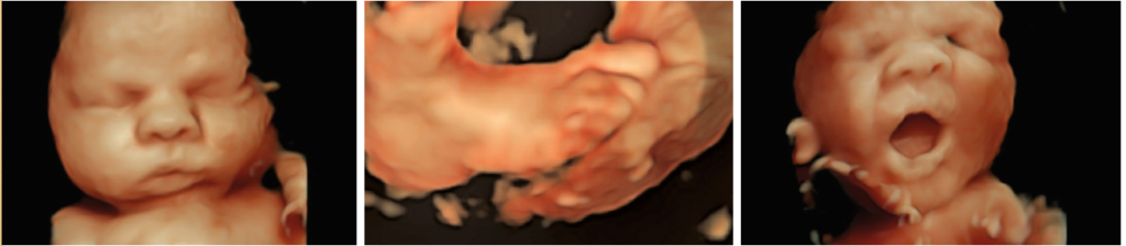
With 3D ultrasound, we can generate frozen images of the surfaces. With 4D ultrasound, we can sometimes see the structures moving through the amniotic fluid, and sometimes the pictures will look very much like a newborn baby.
3D sonography is not part of the standard obstetric ultrasound scan; however, in some specific situations, 3D sonography can be very useful, particularly when we identify an abnormality of the face, brain or spine.
It is important to realize that 3D ultrasound can be very misleading and sometimes is very frightening. We have had many women referred to us who had a 3D ultrasound examination where the baby appeared to have a major abnormality, that was in fact not an abnormality at all, but an irregularity in the image due to the way in which the computer puts together two-dimensional images into a three-dimensional picture. (The same computer technology that was used to create Jar Jar Binks and his friends in Star Wars is used in 3D sonography.)
It is also important to realize that, in many cases, the baby’s position will prevent us from being able to obtain three-dimensional images of a specific structure. For good three-dimensional images, we have to start out with clear two-dimensional images and a surface that is outlined by amniotic fluid.